
Cruciate ligament repair · Sevilla Veterinary Hospital, Santa Monica
TPLO Surgery for Dogs — Cruciate Ligament (ACL) & Knee Surgery Techniques
TPLO vs TTA vs Lateral Suture — how the technique is chosen
TPLO, TTA and lateral suture: procedure, implants, suitability and recovery
| Feature | TPLO | TTA | Lateral Suture |
|---|---|---|---|
| What it does | Cuts and rotates the upper end of the tibia (the tibial plateau) so the plateau is level; the knee is stable through bone geometry. | Cuts and advances the front of the tibia so the patellar tendon neutralizes forward thrust. | A strong suture outside the joint holds the tibia in place while fibrous tissue forms and takes over. |
| Bone cut (osteotomy) | Yes — radial cut through the tibial plateau | Yes — cut through the tibial crest | No |
| Implants | Plate and screws | Titanium cage, plate and screws; bone graft in the gap | Synthetic suture with bone anchor/tunnel |
| Dog weight | Any size; recommended for medium, large and giant breeds | Medium and large dogs with suitable tibial anatomy | Generally dogs under about 30 lb |
| Activity level | Active, working and athletic dogs | Moderately active dogs | Less active or older dogs |
| Tibial plateau angle | Corrected to roughly 4–10°; suits steep plateaus | Depends on plateau slope and tibial shape seen on x-ray | Not altered |
| Restricted activity | About 2–3 months total rehabilitation | About 6–8 weeks | About 6–8 weeks |
| Relative cost | Higher — specialized implants and longer surgery | Higher — specialized implants and longer surgery | Lowest of the three |
No single technique is right for every dog. At consultation Dr. Pedraza reviews the radiographs, examines the knee and discusses your dog's weight, age, activity level and any other orthopedic issues before recommending a technique. The goal is the same in every case: a stable, comfortable knee and a return to normal life.
When does a dog need cruciate ligament surgery?
The cranial cruciate ligament (CCL) sits inside the knee (stifle) joint and stops the tibia from sliding forward under the femur every time the dog puts weight on the leg.
Injury to the cranial cruciate ligament (the dog's equivalent of the human ACL) is one of the most common orthopedic injuries in dogs. This page explains, in plain language, the techniques used most often (CBLO is covered under Other techniques): TPLO, TTA and lateral suture (extracapsular) repair. Dr. Antonio Pedraza Reales explains when each one is recommended, what happens during knee surgery, and what recovery looks like at home. If you already have a diagnosis and radiographs, you can request a free surgical consultation and receive a flat, all-inclusive written quote.
In most dogs the ligament does not tear in a single dramatic accident; it degenerates gradually and then gives way, often during ordinary activity. Genetics, body weight, conformation and age all contribute. Because the process is degenerative, a meaningful proportion of dogs go on to rupture the ligament in the other knee as well.
Once the ligament has failed, the knee is unstable. Every step lets the tibia shift forward (a movement called cranial tibial thrust), which grinds the joint surfaces, damages the meniscus and drives progressive arthritis. For most dogs — and for essentially all medium and large dogs — surgery is the treatment that reliably restores comfortable, long-term use of the leg. Braces, medication, supplements and physical therapy can help manage pain and support recovery, but they do not correct the instability itself. The earlier the joint is stabilized, the less arthritis and meniscal damage accumulate.
Three ways a cruciate rupture presents
- Partial rupture. The ligament is frayed but not completely torn. The dog is intermittently lame, often worse after exercise or after rest, and may seem to "improve" for weeks before flaring again. Partial tears almost always progress to complete tears.
- Complete (chronic) rupture. The ligament has given way after months of degeneration. The lameness is persistent, the dog may toe-touch or hold the leg up, muscle mass on the affected thigh shrinks, and the knee often feels thickened on the inside from scar tissue.
- Traumatic rupture. Less common: a sudden tear in an otherwise healthy ligament after a fall, a slip on a hard floor or a hard twist during play. Lameness is acute and often non-weight-bearing.
Whatever the presentation, the typical signs are: hind-limb lameness, stiffness after rest, sitting with the affected leg stretched out to the side, reluctance to jump or climb stairs, and swelling on the inside of the knee.
How the diagnosis is made
Diagnosis begins with an orthopedic examination of the stifle — the dog's knee joint. The veterinarian feels for the two classic signs of instability — cranial drawer and tibial thrust — and checks for joint effusion and pain. Because a nervous dog tenses the leg, the examination is frequently repeated under sedation.
Radiographs (x-rays) are then taken in standardized positions. They confirm joint effusion and arthritic change, rule out other causes of lameness, and — critically for surgical planning — allow measurement of the tibial plateau angle: the slope of the upper end of the tibia (the tibial plateau) relative to its long axis. In TPLO this measurement determines exactly how far the bone segment must be rotated so that the post-operative plateau sits in the target range of roughly 4 to 10 degrees, where the forward thrust is neutralized.
At surgery, the joint is inspected and the meniscus — the cartilage cushion between femur and tibia — is examined. A torn or crushed portion of meniscus is removed; healthy meniscus is left in place. Untreated meniscal damage is a common cause of ongoing pain after an otherwise successful repair, so this assessment is part of every cruciate procedure we perform.
TPLO (Tibial Plateau Leveling Osteotomy)
What the surgery does
TPLO does not repair or replace the torn ligament.
Instead, it changes the geometry of the knee so the ligament is no longer needed to keep the joint stable.
In a normal dog the tibial plateau slopes backward; that slope is what makes the tibia slide forward when the ligament fails. TPLO cuts the upper end of the tibia (the tibial plateau), rotates the plateau until it is nearly level, and fixes it in the new position with a bone plate and screws. With the plateau level, weight-bearing itself presses the femur straight down onto the tibia and the instability disappears.
TPLO is the technique Dr. Pedraza performs most often. Because stability comes from bone geometry rather than from an implanted band or the dog's own scar tissue, it holds up under sustained heavy loading and provides the most predictable long-term function in larger patients.
Step by step
- Planning. Pre-operative radiographs are used to measure the tibial plateau angle and select the plate size and the rotation required to bring the plateau into the 4–10° target range.
- Anesthesia and preparation. The dog is placed under general anesthesia with continuous monitoring; the leg is clipped and prepared aseptically. Pain control is started before the first incision and continued throughout.
- Joint inspection. The stifle is examined, the remnants of the torn ligament are removed and the meniscus is assessed. Damaged meniscal tissue is excised.
- Osteotomy. A curved (radial) cut is made through the upper end of the tibia (the tibial plateau) with a specially shaped saw blade.
- Rotation. The plateau segment is rotated by the pre-calculated amount and held temporarily in position.
- Fixation. A TPLO plate and screws are applied to the inside of the tibia to hold the bone segments while they heal together. Modern locking plates give a rigid construct that is well tolerated and rarely needs to be removed.
- Closure and radiographs. The incision is closed in layers and post-operative x-rays confirm the plateau angle and implant position.
Most dogs go home within a day of surgery with written discharge instructions, pain medication and, where indicated, a short course of antibiotics.
Recovery after TPLO
The bone cut typically heals over about 8 weeks, but full rehabilitation and return to unrestricted activity takes about 2 to 3 months. Many dogs bear weight on the leg within days; that early comfort is exactly why strict confinement matters — the plate holds the bone, but the bone itself is still healing. The detailed home-care protocol is set out under Aftercare and recovery below.
TTA (Tibial Tuberosity Advancement)
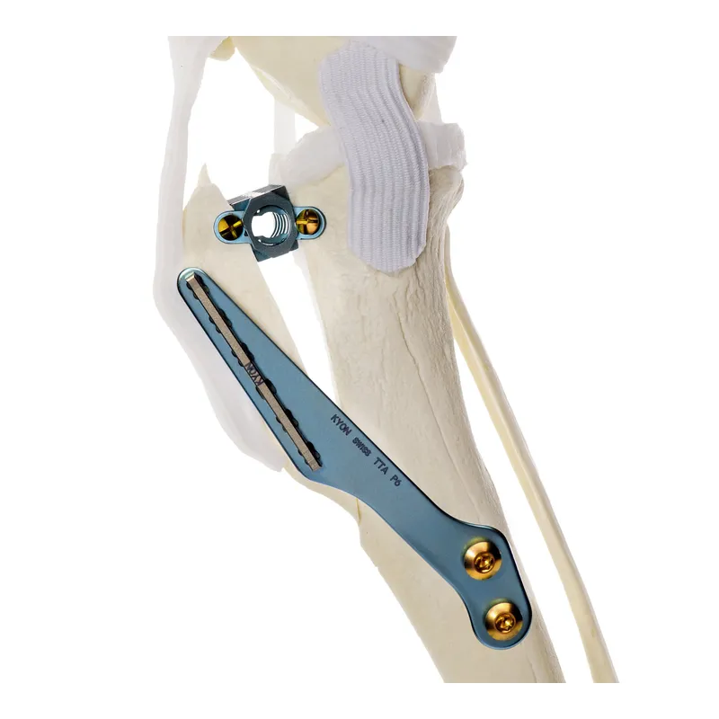
How TTA differs from TPLO
TTA addresses the same problem — forward thrust of the tibia in a knee without a functioning cruciate ligament — from a different angle.
Rather than rotating the tibial plateau, TTA cuts the front of the tibia (the tibial tuberosity, where the patellar tendon attaches) and moves it forward. Advancing the tuberosity changes the direction in which the patellar tendon pulls so that, under load, it becomes perpendicular to the tibial plateau and the forward shear force is neutralized.
Both procedures are "osteotomy" techniques: they rely on cutting and re-positioning bone, then letting it heal in a new geometry. TTA is generally considered less invasive to the weight-bearing part of the tibia, because the cut is made in the front, non-load-bearing crest. It is a good option for certain dogs depending on the individual anatomy — in particular the steepness of the tibial plateau and the shape of the tibia — which is assessed on the pre-operative radiographs. Dr. Pedraza will explain at consultation why TPLO or TTA is the better fit for a specific dog.
Implants and bone graft
The advanced tuberosity is held in its new position with a spacer (a "cage") and a plate with screws or a fork. Dr. Pedraza's preference for TTA is titanium implants: titanium is light, highly biocompatible and well tolerated by bone and soft tissue over the long term. The gap created by moving the tuberosity forward is packed with bone graft, which encourages new bone to bridge the space and helps the osteotomy heal more quickly.
Recovery after TTA
Recovery from TTA is broadly similar to TPLO. Most dogs are comfortable on the leg within days, and activity is restricted for about 6 to 8 weeks while the bone heals, with radiographs taken toward the end of that period before normal exercise is reintroduced. Home care follows the same protocol described under Aftercare and recovery.
Lateral Suture / Extracapsular Technique
How it stabilizes the knee
The lateral suture — also called the extracapsular repair or "fishing-line" technique — is the traditional method of cruciate stabilization and does not involve cutting bone.
A strong synthetic suture is anchored around the small bone behind the knee (the lateral fabella) and through a tunnel in the front of the tibia, running outside the joint capsule in roughly the same direction as the original ligament. Tightened, it stops the tibia from sliding forward.
The suture is not intended to last for the life of the dog. Over the following weeks the joint capsule and surrounding tissues thicken and lay down fibrous (scar) tissue in response to the surgery; it is this periarticular fibrosis that gives the knee its long-term stability. In time the suture itself stretches or breaks — that is expected, and by then the fibrous tissue has taken over its job.
Which dogs it suits
Because it depends on scar tissue rather than on bone geometry, the lateral suture is most suitable for dogs under about 30 pounds and in less active or older patients, where the forces on the knee are smaller. In larger, heavier or athletic dogs the suture is more likely to stretch or fail before adequate fibrous tissue has formed, and results are less consistent than with TPLO or TTA. That is why we generally recommend an osteotomy technique for dogs above that weight.
The meniscus is inspected and damaged tissue removed exactly as in the other techniques. Recovery follows a similar restricted-activity period of 6 to 8 weeks, and physical therapy is especially helpful because muscle strength and controlled loading encourage the fibrous tissue to mature.
Is there a cost difference?
The lateral suture uses simpler implants and a shorter surgical time than TPLO or TTA, so it is normally the least expensive of the three. The osteotomy techniques require specialized plates, screws and instrumentation and a longer, more technically demanding procedure. That said, the technique is chosen on what is right for the dog — weight, activity, age and the tibial plateau angle — not on price. After a free surgical consultation we provide a flat, all-inclusive written quote for the recommended procedure covering the surgery, anesthesia, pain management, hospitalization, medications to go home, and the scheduled post-operative rechecks including the follow-up X-ray, so there are no surprise add-ons. Contact us to arrange a consultation.
Aftercare and recovery — what to expect at home
Surgery is half of the outcome; the other half happens at home in the weeks that follow. Whatever the technique, the principles are the same.
Rehabilitation window.
Plan for 2 to 3 months of rehabilitation after TPLO and about 6 to 8 weeks of restricted activity after TTA or lateral suture. Your written discharge instructions give the schedule for your dog.
Strict exercise restriction. No running, jumping, stairs, rough play or off-leash time until Dr. Pedraza clears it. Most complications after cruciate surgery come from too much activity too soon, especially once the dog feels better.
Crate rest / confinement.
When you cannot directly supervise, your dog should be in a crate or a small room with non-slip flooring. Block access to furniture and stairs.
Leash at all times outdoors. Bathroom breaks are on a short leash only, for the first several weeks — even in a fenced yard.
Harness and support. A harness (rather than a collar) gives you better control, and a sling or towel under the belly can help a larger dog on slippery surfaces or steps during the first days.
Incision care.
Check the incision daily for redness, swelling or discharge. Prevent licking with an e-collar or protective suit. Sutures or staples are removed within about two weeks of surgery.
Medications. Give the prescribed pain relief and anti-inflammatory medication on schedule, and finish any course of antibiotics as directed. Do not give human pain medicines.
Physical therapy.
Gentle, controlled physical therapy — passive range-of-motion, short slow leash walks that increase gradually, and physical therapy with a rehabilitation provider, if recommended — rebuilds muscle and helps the joint move well. We will tell you when and how to begin. See also our other orthopedic services.
Weight management. Keeping your dog lean reduces the load on both knees and lowers the chance of problems in the opposite leg.
Radiographic recheck. Follow-up x-rays are taken at around 6 to 8 weeks to confirm the bone is healing and the implants are stable. Only after that check is activity gradually increased toward normal.
Call us if: the dog suddenly stops using the leg after having used it, the incision opens or discharges, there is a fever, or pain seems to be getting worse rather than better.
Other techniques you may read about
Other techniques you may read about
CBLO (CORA-Based Leveling Osteotomy) is a more recent leveling osteotomy that, like TPLO, changes the angle of the tibial plateau so the knee no longer depends on the torn ligament. Dr. Pedraza performs CBLO for selected dogs; whether it is preferable to TPLO for your dog is decided at the consultation from the X-rays.
You may also read about MMP (Modified Maquet Procedure), a variant of TTA. Ask us at the consultation which technique is appropriate for your dog.
For a second opinion, ask us to review the case.
Frequently asked questions about TPLO
What is a TPLO surgery?
What is a TPLO surgery?
TPLO stands for Tibial Plateau Leveling Osteotomy. It is a surgery for dogs with a torn cranial cruciate ligament (the canine ACL). Instead of replacing the ligament, the upper end of the tibia (the tibial plateau) is cut, rotated until it is nearly level, and fixed with a plate and screws. Leveling the plateau stops the tibia from sliding forward, so the knee is stable even without the ligament.
How is the TPLO procedure done on a dog?
How is the TPLO procedure done on a dog?
Under general anesthesia the knee is inspected and any damaged meniscus is removed. A curved cut is made through the upper end of the tibia (the tibial plateau), the plateau segment is rotated by an amount calculated from pre-operative x-rays, and a bone plate is applied to hold it while the bone heals. Post-operative x-rays confirm the new angle. Most dogs go home within a day of surgery.
What is the TPLO recovery time?
What is the TPLO recovery time?
Most dogs put weight on the leg within days, but the bone needs roughly 8 weeks to heal and full rehabilitation takes about 2 to 3 months. During that time activity is strictly restricted, with crate rest, leash-only bathroom breaks and gradually increasing controlled walks. A radiographic recheck at around 6 to 8 weeks confirms healing before normal activity resumes.
TPLO vs TTA — what is the difference?
TPLO vs TTA — what is the difference?
Both are osteotomy techniques that neutralize the forward thrust of the tibia. TPLO rotates the tibial plateau to level it; TTA moves the front of the tibia forward so the patellar tendon does the stabilizing. Which one suits a particular dog depends on the slope of the tibial plateau, the shape of the tibia, and the dog's weight and activity, all assessed on the pre-operative radiographs. Recovery timelines are similar.
What affects the cost of TPLO surgery?
What affects the cost of TPLO surgery?
The main factors are the size of the dog (implant size, anesthesia and medication doses), whether one or both knees need surgery, the extent of meniscal damage, the technique chosen (TPLO and TTA use specialized implants and take longer than the lateral suture), and what is included in the quote. We do not publish a price list because every case is different; instead, after a free surgical consultation you receive a single flat, all-inclusive written quote with no hidden extras.
TPLO, TLPO or TPO?
TPLO, TLPO or TPO?
The correct term is TPLO — Tibial Plateau Leveling Osteotomy. 'TLPO' and 'TPO surgery' are common misspellings of the same procedure.
Can a small dog have TPLO surgery?
Can a small dog have TPLO surgery?
Yes. TPLO can be performed on small dogs using appropriately sized plates, and it is often recommended for small dogs that are very active. For less active dogs under about 30 pounds the lateral suture (extracapsular) technique is also a reasonable option. Dr. Pedraza discusses both at consultation and recommends based on the individual dog rather than size alone.
When is surgery needed for a torn cruciate ligament?
When is surgery needed for a torn cruciate ligament?
Surgery is recommended when the knee is unstable — which is the case in essentially all complete ruptures and most partial ruptures that are causing lameness. Medication and rest can ease the pain for a while, but they do not correct the instability, and the longer an unstable knee is left, the more arthritis and meniscal damage develop. The exception is a very small, low-activity dog where conservative management may be trialled first; that decision is made together at consultation.
Not sure which technique is right for your dog?
Next step
Bring your dog's records and radiographs, or have your veterinarian send them to us, and Dr. Pedraza will examine the knee and recommend the procedure that fits. The surgical consultation is free, and you leave with a flat, all-inclusive written quote.